Gluconeogenesis on Keto: Protein, Glucose, and Ketosis
Gluconeogenesis is the regulated production of glucose from lactate, glycerol, and glucogenic amino acids. In humans, the liver and renal cortex are the principal sources of newly made glucose released into blood. The pathway responds to hormones, substrate delivery, cellular energy state, and acid-base balance. It is not a gram-for-gram conversion of dietary protein, but it is also not governed by "demand" alone.1
Red blood cells require glucose because they lack mitochondria. During prolonged fasting or carbohydrate restriction, the brain can replace part, not all, of its glucose use with ketone bodies. Gluconeogenesis therefore continues while ketone production rises.1
Reading evidence in context
- Observed outcome
- Certainty
- Duration
- What remains unknown
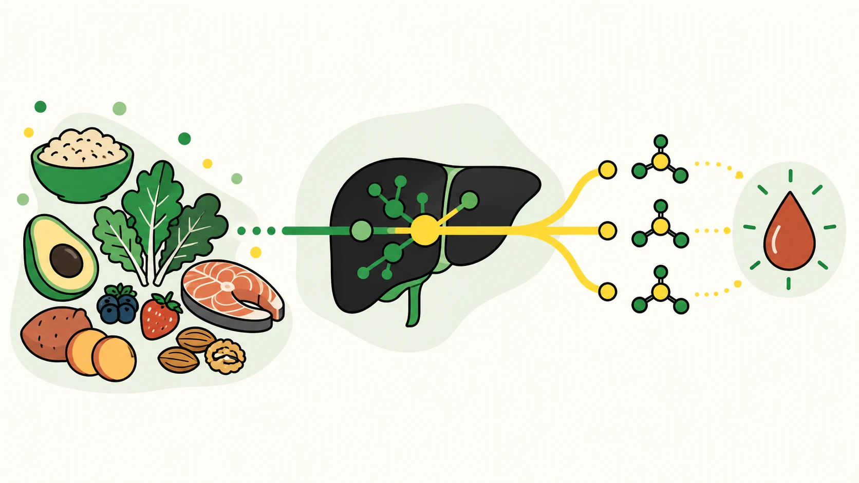
What goes into gluconeogenesis?
The pathway starts with carbon that is already circulating or released from stored nutrients. Its major precursors are:
| Precursor | Main source | How it contributes |
|---|---|---|
| Lactate | Red blood cells and working tissues | Returns to the liver and kidney for conversion through the Cori cycle |
| Glycerol | Breakdown of triglycerides | Enters the pathway through a three-carbon intermediate |
| Glucogenic amino acids | Dietary protein and normal protein turnover | Enter through pyruvate or citric-acid-cycle intermediates |
| Pyruvate and related intermediates | Several metabolic pathways | Supply carbon for glucose production |
Gluconeogenesis is not simply glycolysis run backward. Three energetically unfavorable glycolysis reactions require bypass enzymes, including phosphoenolpyruvate carboxykinase and glucose-6-phosphatase. The pathway also consumes energy, so its rate depends on cellular energy and redox state as well as available carbon.1
Which organs make glucose?
The liver is the main fasting glucose supplier through both glycogen breakdown and gluconeogenesis. Renal gluconeogenesis occurs mainly in proximal tubules of the renal cortex and becomes more important with prolonged fasting and metabolic acidosis. The small intestine expresses the required enzymes and may contribute under some conditions, but its quantitative contribution to circulating glucose in humans is less certain.1 2
Human measurements illustrate why a fixed organ percentage would be misleading. In an arterial-venous isotope study, Ekberg and colleagues estimated that the kidney supplied about 5% of endogenous glucose after an overnight fast and about 24% after 60 hours of fasting. Other tracer methods have produced different estimates, but the direction is consistent: the renal contribution changes with nutritional and metabolic conditions.3
The kidney also links glucose production to acid-base control. During acidosis, proximal-tubule metabolism of glutamine supports ammonium excretion and bicarbonate generation while supplying carbon for glucose production. This is one reason renal regulation cannot be reduced to the same hormonal summary used for the liver.2
How is the pathway regulated?
Insulin suppresses hepatic glucose production, while glucagon and other counter-regulatory signals support it during fasting. Transcription of pathway enzymes, allosteric regulation, precursor delivery, fatty-acid oxidation, and the cell's ATP and NADH balance also affect the rate. Renal regulation overlaps with hepatic regulation but is not identical, particularly during acidosis.1
Reducing gluconeogenesis to either precursor supply or glucose demand creates a false binary. More precursor does not guarantee an equal rise in glucose released to blood. At the same time, substrate availability can affect flux, and gluconeogenesis can be inappropriately elevated in insulin resistance and diabetes. "Regulated, not gram-for-gram" is the more accurate conclusion.
During an overnight fast, liver glycogen breakdown and gluconeogenesis both support circulating glucose. As fasting continues and liver glycogen falls, the share supplied by gluconeogenesis rises. Ketogenesis also increases, so the two pathways operate together rather than acting as opposing metabolic switches. The science of ketosis explains that parallel response.
What do protein-feeding studies actually show?
Three frequently cited human studies answer different questions. None was designed to identify a protein dose that keeps every person in nutritional ketosis.
| Study | Design and result | What it cannot establish |
|---|---|---|
| Fromentin et al., 2013 | Eight healthy adults ate one meal containing 23 g of intrinsically labeled egg protein after an overnight fast. Over eight hours, total glucose production was 50.4 ± 7.7 g, and 3.9 ± 0.7 g came from the meal's amino acids. Total endogenous glucose production remained stable for six hours and then fell.4 | The study did not test keto-adapted participants, a high protein dose, blood beta-hydroxybutyrate (BHB), or whether anyone left ketosis. It did not show that liver glycogen supplied all remaining glucose. |
| Veldhorst et al., 2009 | Ten healthy men completed short high-protein, carbohydrate-free and normal-protein, carbohydrate-containing conditions after exhaustive exercise. Fractional gluconeogenesis was higher in the high-protein condition, while endogenous glucose production was lower. The difference in absolute gluconeogenesis did not reach conventional statistical significance.5 | The 1.5-day intervention and glycogen-lowering exercise protocol do not define a usual keto response or a protein threshold. |
| Linn et al., 2000 | Nine habitual high-protein consumers were compared with nine matched normal-protein consumers. The high-protein group had higher fractional gluconeogenesis, fasting glucagon, and modestly higher endogenous glucose production under low-insulin conditions.6 | This small observational comparison was not randomized and did not study a ketogenic diet or BHB response. |
The Fromentin experiment shows why dietary amino acids should not be counted as if every gram became glucose. The Veldhorst and Linn results show why the opposite absolute claim is also unwarranted: protein intake, adaptation, hormonal state, and study conditions can change measured flux.
These studies rule out a simple one-to-one conversion. They do not show that protein never changes gluconeogenesis or ketone concentration, and they do not define a protein dose that preserves ketosis for everyone.
What does this mean for protein on keto?
Protein has functions that cannot be replaced by fat. Amino acids support tissue maintenance, enzymes, transport proteins, and immune functions. After a meal, amino acids can be incorporated into protein, oxidized, converted into other metabolites, or used as gluconeogenic precursors. The proportions vary with the meal and the person's physiology.
There is no controlled human dose-response study that supplies a universal protein cutoff for "staying in ketosis." Protein needs also differ with body size, age, energy intake, training, illness, and clinical goals. General protein recommendations and therapeutic ketogenic-diet prescriptions answer different questions; neither should be presented as a biochemical ceiling derived from the tracer studies above.
Use the macro calculation guide to understand how protein can be planned alongside carbohydrate and energy intake. The high-protein, low-carb guide covers the separate evidence on higher-protein diets. People with kidney disease or a medically prescribed ketogenic diet need an individualized target from their clinical team.
Can a ketone reading reveal gluconeogenesis?
A change in blood BHB after a meal cannot identify gluconeogenesis as the cause. The reading reflects ketone production, tissue use, distribution, and clearance. Recent exercise, fasting duration, energy intake, alcohol, illness, medications, and test variation can also change it.
The glucose-ketone index (GKI) was proposed for research on metabolic therapy in brain cancer. It has since been used in some therapeutic-diet studies, but it has no validated target for general wellness, fat loss, or diagnosing a protein response.7 See the GKI evidence guide before interpreting the ratio.
If testing is required for diabetes or a prescribed ketogenic therapy, use the thresholds and schedule provided by the treating team. Do not reduce insulin or other medication in response to a diet or home ketone reading without clinical guidance. The ketosis and ketoacidosis guide explains why glucose and ketone readings alone cannot assess blood acidity.
Frequently Asked Questions
Does dietary protein turn into glucose?
Is gluconeogenesis controlled only by the body's glucose demand?
Can research identify one protein limit for ketosis?
Do gluconeogenesis and ketogenesis happen at the same time?
Works cited
Article history
- Consolidated the retired glossary entry; rewrote organ-specific physiology and the limits of protein-tracer evidence
- First published
Your learning guide